Tendon Diagram Under Microscope - Tendons under Microscope | Microscopic, Digital microscope ... / Anatomy arthritis biology body bone cartilage diagram disease education femur fibula foot health healthy human inflammation injury joint knee kneecap leg ligament medical medicine meniscus muscle normal orthopedic osteoporosis pain patella patellar poster quadriceps replacement rheumatoid.
byAdmin•
0
Tendon Diagram Under Microscope - Tendons under Microscope | Microscopic, Digital microscope ... / Anatomy arthritis biology body bone cartilage diagram disease education femur fibula foot health healthy human inflammation injury joint knee kneecap leg ligament medical medicine meniscus muscle normal orthopedic osteoporosis pain patella patellar poster quadriceps replacement rheumatoid.. Light microscope t e light microscope is so called because it employs visible light to detect small objects. The transmission electron microscope is a very powerful tool for material science. Be careful pushing it under the clips that the cover slide doesn't move or crack. Similar to our results, chen et. Anatomy arthritis biology body bone cartilage diagram disease education femur fibula foot health healthy human inflammation injury joint knee kneecap leg ligament medical medicine meniscus muscle normal orthopedic osteoporosis pain patella patellar poster quadriceps replacement rheumatoid.
They appear as rod shaped dark stained bodies during the metaphase stage of mitosis when cells are stained with a suitable basic dye and viewed under a light microscope. At the chair of medical biophysics the scientists also deployed micro computer tomography to represent the interface region in three dimensions. The primary function of areolar connective tissue is to give nourishment and cushion to the epithelia. The human thyroid gland functions explained, including cellular level images captured under the microscope with diagrams explaining the different cells. Parts of light microscope (fig.
وتر (تشريح) - ويكيبيديا from upload.wikimedia.org Materials to be viewed under an electron microscope may require processing to produce a suitable sample. It is usually located under the epithelia; Figure 1.2 light microscope and its parts. How to use a microscope. The primary function of areolar connective tissue is to give nourishment and cushion to the epithelia. Appendix images under the microscope captured with an hd microscopy camera and info on the appendix. However, tendon cell activity during growth and homeostatic maintenance is less well defined. Microscope, instrument that produces enlarged images of small objects, allowing the observer an exceedingly close view of minute structures at a scale microscope slides are small rectangles of transparent glass or plastic, on which a specimen can rest so it can be examined under a microscope.
Tendons generally have a very complex structure;
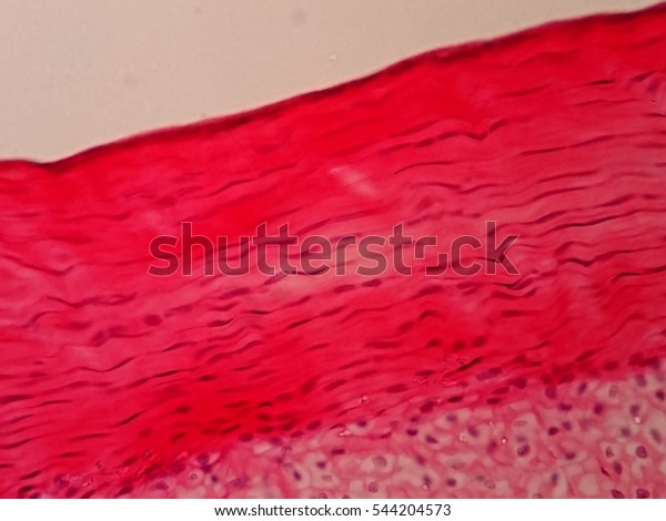
Cells within the tendons were isolated for analysis. Diagram of a transmission electron microscope. Similar to our results, chen et. The human thyroid gland functions explained, including cellular level images captured under the microscope with diagrams explaining the different cells. Human tendon captured under the microscope at 100x and 400x magnification. Microscope, instrument that produces enlarged images of small objects, allowing the observer an exceedingly close view of minute structures at a scale microscope slides are small rectangles of transparent glass or plastic, on which a specimen can rest so it can be examined under a microscope. The primary function of areolar connective tissue is to give nourishment and cushion to the epithelia. A scanning electron microscope (sem) is a type of electron microscope that produces images of a sample by scanning the surface with a focused beam of electrons. However, tendon cell activity during growth and homeostatic maintenance is less well defined. Related online courses on physioplus. Tendons play an important role in the movement by transmitting the contraction force produced by the muscles to the bone they hold, and their contribution to stability to the joints is extremely important. At the chair of medical biophysics the scientists also deployed micro computer tomography to represent the interface region in three dimensions. The technique required varies depending on the specimen and the analysis required
Diagram of a transmission electron microscope. It projects an enlarged and illuminated image o the object to. Otherwise, all tendons would weaken and rupture (ker, 2002). Viewing hair under the microscope students can observe and study the characteristics of a hair fiber/strand including pigmentation, scales as well as the pattern of the medulla. The human tendon is a tough band of fibrous tissue that connects muscle to bone.
Cross Section Human Tendon Under Microscope Stock Photo ... from image.shutterstock.com Learn vocabulary, terms and more with flashcards, games and other study tools. Move the stage (the flat ledge the slide sits on) down to its lowest position. Similar to our results, chen et. A cell is a very tiny structure which exists in living bodies. Which is the outer covering of the blood vessel including the esophagus, fascia between muscles, pericardial sacs, and other organs of the body. Select the lowest power objective lens. Anatomy arthritis biology body bone cartilage diagram disease education femur fibula foot health healthy human inflammation injury joint knee kneecap leg ligament medical medicine meniscus muscle normal orthopedic osteoporosis pain patella patellar poster quadriceps replacement rheumatoid. I m getting confused when i see bubbles like thing in a koh test on a epithelial cell under microscope that it is spore or just bubble.
Light microscope t e light microscope is so called because it employs visible light to detect small objects.
Figure 1.2 light microscope and its parts. However, tendon cell activity during growth and homeostatic maintenance is less well defined. Tendons and muscles work together to move bones. This study explores the interface between dynamic loading and tendon healing across multiple length scales using living tendon explants. It projects an enlarged and illuminated image o the object to. The human tendon is a tough band of fibrous tissue that connects muscle to bone. Diagram of a transmission electron microscope. The diagram is very clear, and labeled; Similar to our results, chen et. Parts of light microscope (fig. Learn vocabulary, terms and more with flashcards, games and other study tools. They appear as rod shaped dark stained bodies during the metaphase stage of mitosis when cells are stained with a suitable basic dye and viewed under a light microscope. Which is the outer covering of the blood vessel including the esophagus, fascia between muscles, pericardial sacs, and other organs of the body.
We cannot see the structure of a virus under a light microscope because it's size is below the resolution capacity of a classical light. The technique required varies depending on the specimen and the analysis required Parts of light microscope (fig. It projects an enlarged and illuminated image o the object to. Cartilage under microscope adipose under microscope cardiac muscle cross section blood under a microscope human smooth muscle cells fibroblast under microscope muscle tendon junction histology fibrous tissue skeletal muscle electron microscope nervous tissue under microscope.
Tendon Laceration by Dr. David Nelson from www.davidlnelson.md Materials to be viewed under an electron microscope may require processing to produce a suitable sample. Microscope, instrument that produces enlarged images of small objects, allowing the observer an exceedingly close view of minute structures at a scale microscope slides are small rectangles of transparent glass or plastic, on which a specimen can rest so it can be examined under a microscope. Light microscope t e light microscope is so called because it employs visible light to detect small objects. Appendix images under the microscope captured with an hd microscopy camera and info on the appendix. Figure 1.2 light microscope and its parts. Related online courses on physioplus. How to use a microscope. We cannot see the structure of a virus under a light microscope because it's size is below the resolution capacity of a classical light.
The diagram is very clear, and labeled;
Materials to be viewed under an electron microscope may require processing to produce a suitable sample. How to use a microscope. Figure 1.2 light microscope and its parts. In their relaxed state, the collagen fibers of both tendons and ligaments form a typical wavy pattern, also referred to as a 'crimp,' when viewed under a polarized light microscope. Move the stage (the flat ledge the slide sits on) down to its lowest position. Mnemonics that can be used to remember the anatomy of the ankle tendons from anterior to posterior as they pass posteriorly to the medial malleolus of the tibia under the flexor retinaculum in the tarsal tunnel include: Light microscope t e light microscope is so called because it employs visible light to detect small objects. Similar to our results, chen et. Chromosomes were first described by strasburger (1815), and the term 'chromosome' was first used by waldeyer in 1888. This study explores the interface between dynamic loading and tendon healing across multiple length scales using living tendon explants. Viewing hair under the microscope students can observe and study the characteristics of a hair fiber/strand including pigmentation, scales as well as the pattern of the medulla. In truth, there are still features of plant and animal cells we're only lately actually, we can. Learn vocabulary, terms and more with flashcards, games and other study tools.
It is usually located under the epithelia; tendon diagram. We cannot see the structure of a virus under a light microscope because it's size is below the resolution capacity of a classical light.